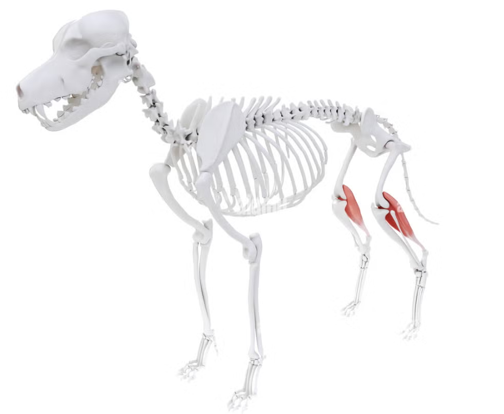
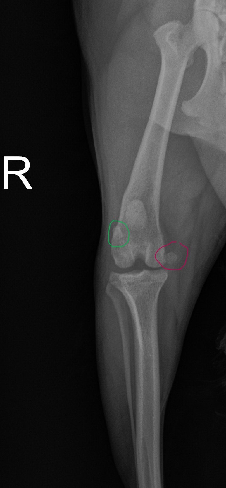
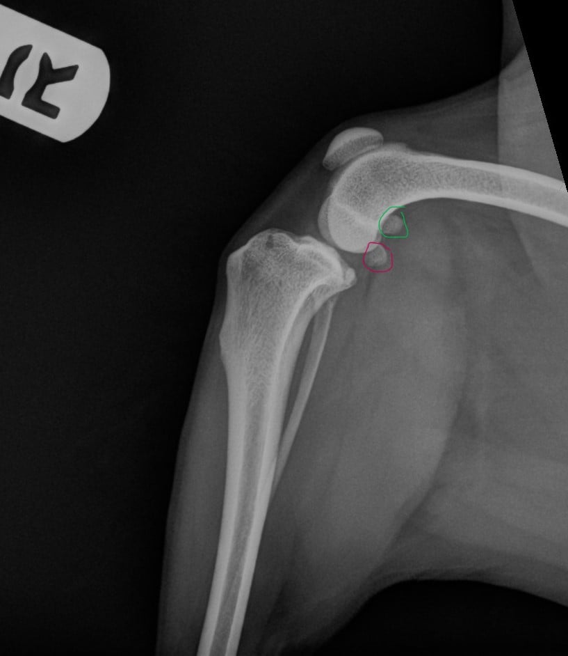
The gastrocnemius muscle is the chief muscle of the calf of the pelvic limb, which flexes the stifle (knee) and hock (ankle). The gastrocnemius muscle has two heads which originate on the distal femur. The medial head of the gastrocnemius originates on the medial aspect of the distal femur and incorporates the medial fabella. The lateral head of the gastrocnemius originates from the lateral aspect of the distal femur and incorporates the lateral fabella. The two heads combine and run distally along the caudal aspect of the tibia to form the common calcaneal (Achilles) tendon.
 Avulsion injuries affecting the origin of the gastrocnemius muscles are rare. Few reports are published regarding the surgical management of these lesions in dogs and cats. Some of these reports are poorly detailed and lack follow-up data, giving surgeons scarce evidence on how to optimally treat these types of lesions. Most lesions are unilateral, acute and traumatic in nature; however, atraumatic bilateral avulsion of the origin of the gastrocnemius has been reported in one dog. Cases present with a history of weight-bearing lameness with different degrees of tarsal hyperflexion, eventually leading to a complete plantigrade stance.

 Avulsion fracture of the medial head or lateral head of the gastrocnemius muscle is a rare injury caused by traction on the origin of the medial head of the muscle on the medial femoral condyle.
Achilles tendon injuries or common calcaneal tendon injuries are most commonly caused by laceration or sharp trauma.
Acute injuries (<48 hours old) to the common calcaneal tendon are most commonly due to lacerations.
Subacute injuries (2-21 days)
Chronic injuries (>21 days). With chronic progressive or intermittent injuries resulting in progressive damage, onset of the disease is often difficult to ascertain.
The severity of the rupture of the tendon has been classified as type I (complete disruption, plantegrade stance), type IIa (musculotendinous rupture, tarsal hyperflexion), type IIb (ruptured tendon but paratenon intact, tarsal hyperflexion), type IIc (gastrocnemius tendon rupture but semitendinosus intact, flexed hock with excessive digit flexion), type III (tendinosis or peritendinitis, normal stance).
Chronic injuries are more problematic to treat and may carry a poorer prognosis since tendon and muscular contracture develops along with fibrous scar tissue at the ruptured tendon ends.